Forschungsprozess
76 Abbildungen aus begutachteter Forschung
Brain imaging studies demonstrate that elevated homocysteine is associated with accelerated rates of brain atrophy, particularly in medial temporal lobe structures critical for memory. B-vitamin treatment appears to slow this atrophy in individuals with elevated baseline homocysteine.
Homocysteine and Dementia: An International Consensus Statement.
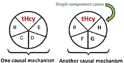
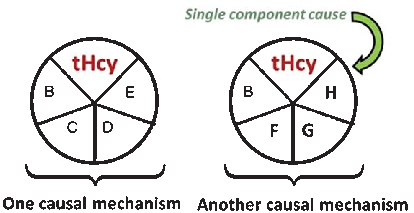
A causal model illustrates how elevated plasma homocysteine may contribute to dementia through multiple pathways, interacting with other risk factors such as age, hypercholesterolemia, and genetic predisposition. No single factor is sufficient alone; rather, combinations of component causes drive disease.
Homocysteine and Dementia: An International Consensus Statement.
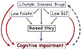
Meta-analyses of cohort studies consistently indicate that elevated homocysteine is associated with approximately doubled risk of Alzheimer's disease. The strength of this association persists after adjustment for common confounders including age, sex, and education.
Homocysteine and Dementia: An International Consensus Statement.
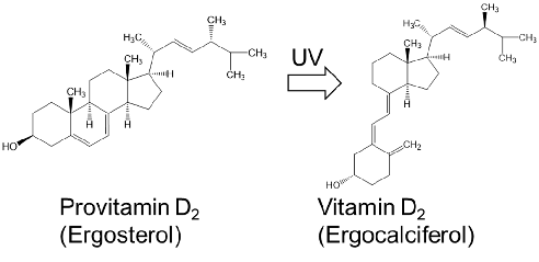
Structures of provitamin D2 and vitamin D2 found in commercially available Chlorella products are displayed, demonstrating the ergosterol-to-ergocalciferol conversion pathway.
Potential of Chlorella as a Dietary Supplement to Promote Human Health.
Growth factor content and cell wall composition of different Chlorella species used in commercial supplements are compared.
Potential of Chlorella as a Dietary Supplement to Promote Human Health.
Safety assessment data for Chlorella supplementation, including heavy metal contamination screening and adverse event reporting from clinical trials.
Potential of Chlorella as a Dietary Supplement to Promote Human Health.
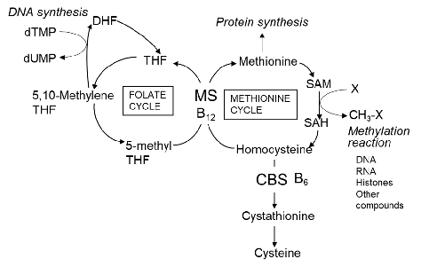
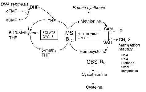
Homocysteine metabolic pathway in mammals is depicted showing how Chlorella-derived folate and vitamin B12 participate as essential cofactors in methionine remethylation and transsulfuration reactions.
Potential of Chlorella as a Dietary Supplement to Promote Human Health.
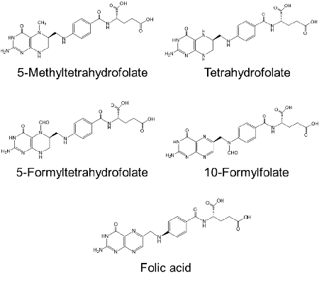
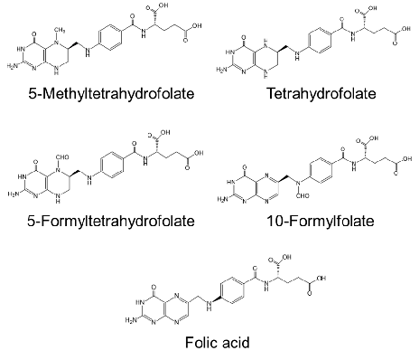
Chemical structures of folate compounds found in commercially available Chlorella products are displayed, including tetrahydrofolate and 5-methyltetrahydrofolate forms.
Potential of Chlorella as a Dietary Supplement to Promote Human Health.
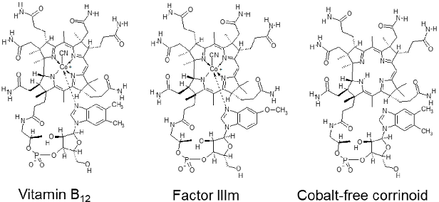
Chemical structures of vitamin B12 and related corrinoid compounds found in Chlorella products are presented, with identification of true cobalamin versus inactive analogues.
Potential of Chlorella as a Dietary Supplement to Promote Human Health.
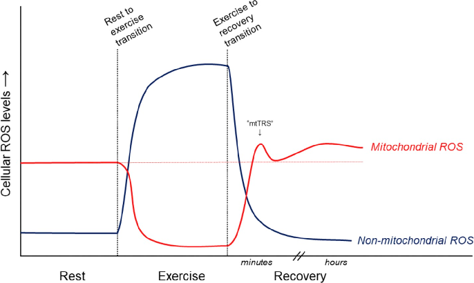
Proposed relative contributions of mitochondrial and non-mitochondrial ROS sources to overall cellular ROS levels in skeletal muscle during and after exercise are depicted. NADPH oxidase, xanthine oxidase, and mitochondrial electron transport chain are the primary generators.
Antioxidant supplements and endurance exercise: Current evidence and mechanistic insights.
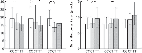
Moderate hyperhomocysteinemia resulting from low folate status is an independent risk factor for cardiovascular disease, dementia, and depression. This figure maps the clinical consequences of impaired folate metabolism due to genetic variants.
Genetic polymorphisms and folate status.
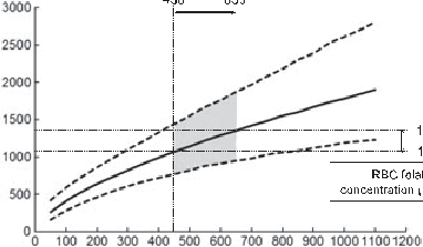
Population-level variation in folate-related genetic polymorphisms influences disease susceptibility across ethnic groups. This figure compares allele frequencies and associated health outcomes for key folate metabolism gene variants.
Genetic polymorphisms and folate status.
![Figure 1. One-carbon metabolism. Abbreviations: PLP, plasma pyridoxal phosphate; MTHFR, methylenetetrahydrofolate reductase; FAD, flavin adenine dinucleotide; FMN, flavin mononucleotide. Adapted from [16].](https://pdfs.citedhealth.com/figures/27854316/97.png)
One-carbon metabolism pathways involving folate, vitamin B12, and vitamin B6 are mapped, showing key enzymatic reactions catalyzed by MTHFR and the roles of FAD and FMN as cofactors in homocysteine recycling.
Causes, Consequences and Public Health Implications of Low B-Vitamin Status in Ageing.
Body composition changes observed in participants following vegan dietary interventions are tracked over time, showing reductions in visceral adiposity and overall fat mass.
Vegan Diet Health Benefits in Metabolic Syndrome.
The complete literature search strategy for this systematic review of vegan diet effects on metabolic syndrome is presented, detailing database queries and Boolean operators used to identify eligible studies.
Vegan Diet Health Benefits in Metabolic Syndrome.
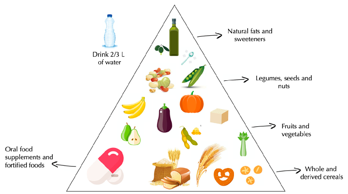
A vegan food pyramid illustrates recommended dietary proportions for plant-based nutrition, organizing food groups by suggested intake frequency to support metabolic health.
Vegan Diet Health Benefits in Metabolic Syndrome.
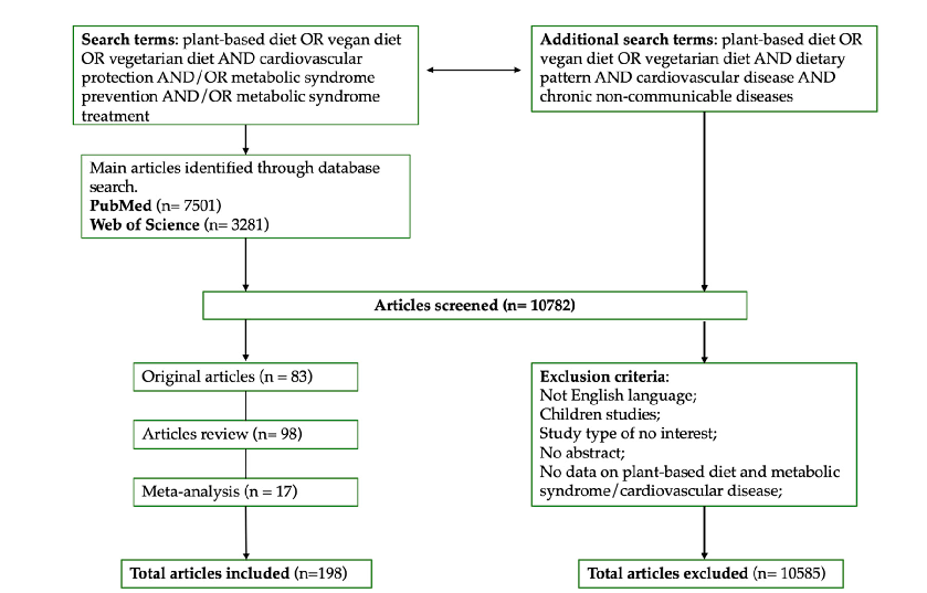
PRISMA flow diagram for study selection in this review of vegan diet and metabolic syndrome is depicted, showing records identified, screened, and ultimately included.
Vegan Diet Health Benefits in Metabolic Syndrome.
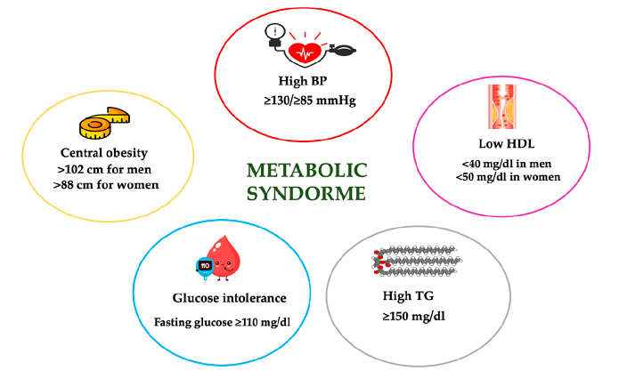
Insulin sensitivity indices before and after vegan dietary intervention are plotted, demonstrating improved glucose homeostasis in the plant-based diet group.
Vegan Diet Health Benefits in Metabolic Syndrome.
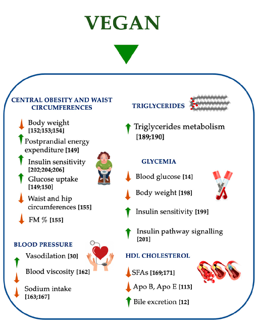
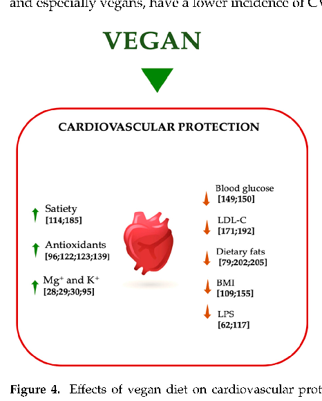
Beneficial effects of a vegan diet on metabolic syndrome components are mapped, showing improvements in apolipoprotein profiles, fat mass reduction, and decreased saturated fatty acid intake alongside their downstream metabolic consequences.
Vegan Diet Health Benefits in Metabolic Syndrome.
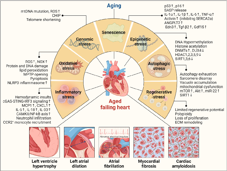
Comprehensive diagram of cardiac aging mechanisms leading to heart failure, depicting how genomic, epigenetic, oxidative, autophagic, and inflammatory stresses drive structural changes including left ventricular hypertrophy and myocardial fibrosis.
Systemic aging fuels heart failure: Molecular mechanisms and therapeutic avenues.
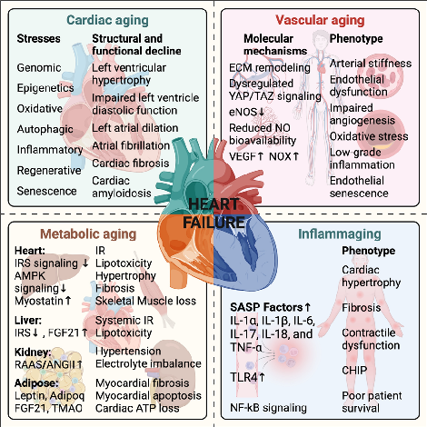
Systemic aging pathways fueling heart failure, summarizing cardiac, vascular, metabolic, and inflammaging mechanisms alongside their associated molecular changes and clinical phenotypes.
Systemic aging fuels heart failure: Molecular mechanisms and therapeutic avenues.
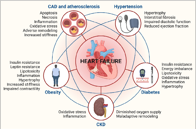
Molecular mechanisms or therapeutic targets related to age-associated heart failure, covering topics such as senescent cell accumulation, RAAS dysregulation, and potential anti-aging interventions.
Systemic aging fuels heart failure: Molecular mechanisms and therapeutic avenues.
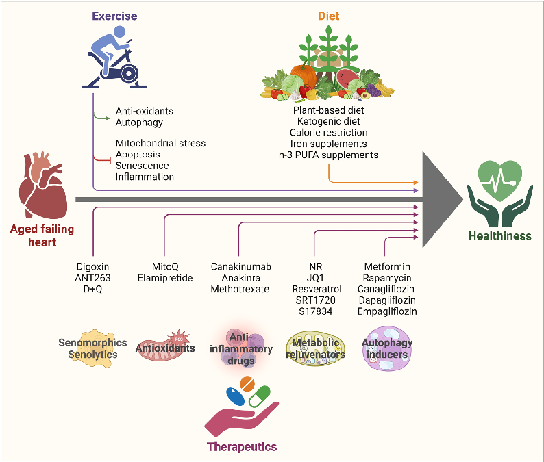
Molecular mechanisms or therapeutic targets related to age-associated heart failure, covering topics such as senescent cell accumulation, RAAS dysregulation, and potential anti-aging interventions.
Systemic aging fuels heart failure: Molecular mechanisms and therapeutic avenues.
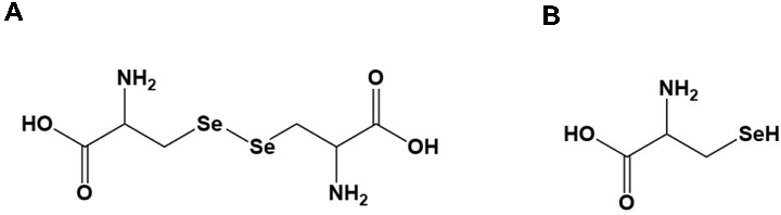
Selenium-Containing Amino Acids Protect Dextran Sulfate Sodium-Induced Colitis via Ameliorating Oxidative Stress …
Seite 1 von 4